A DAN Boater Travel Health and Safety Guide
Boating can be a soothing and relaxing activity, but it also can be a source of body aches and musculoskeletal injuries. Whether from the strenuous activity unique to sailing, the constant muscular adjustments your body makes on a swaying boat or the repetitive motions associated with other water sports popular with boaters, it is not unusual to experience body pains more than those usually associated with “just being tired.” The boat’s movement and structure can cause additional pressure to be exerted on one’s musculature. In addition, slips and falls while boating can cause serious as well as minor musculoskeletal injuries.
This guide addresses some of the musculoskeletal injuries one may experience on a boat and how to manage them.
The following sections of this guide provide a summary of the causes and symptoms of each condition as well as tips for prevention and instructions for treatment.
— Contributors —
James M. Chimiak, MD
Christopher T. Daley, MD
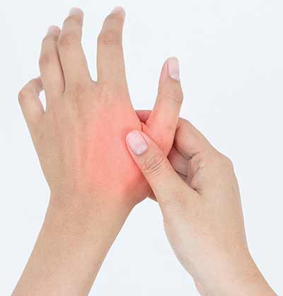
A sprain involves an injury of the soft tissue surrounding a joint where one or more of the supporting ligaments — the strong bands of tissue connecting two bones — become stretched or torn. Common locations for sprains include the ankles, knees, wrists and thumbs.
A sprain should not be confused with a strain, which refers to an injury in which muscle or tendon fibers stretch or tear. A tendon connects the muscle to the bone. Common strains involve the hamstring and lower back
Symptoms of a sprain include pain, swelling and difficulty using the affected joint. Bruising may also occur, but it is usually delayed in appearance and often develops away from the affected joint, as blood tracks along the tissues before surfacing to the skin. A pop or tearing sensation may be experienced at the time of injury.
Generally, the greater the pain and swelling, the more severe the injury. If the ligament supporting the joint is completely torn, a person will often experience instability when attempting to bear weight through the affected joint.
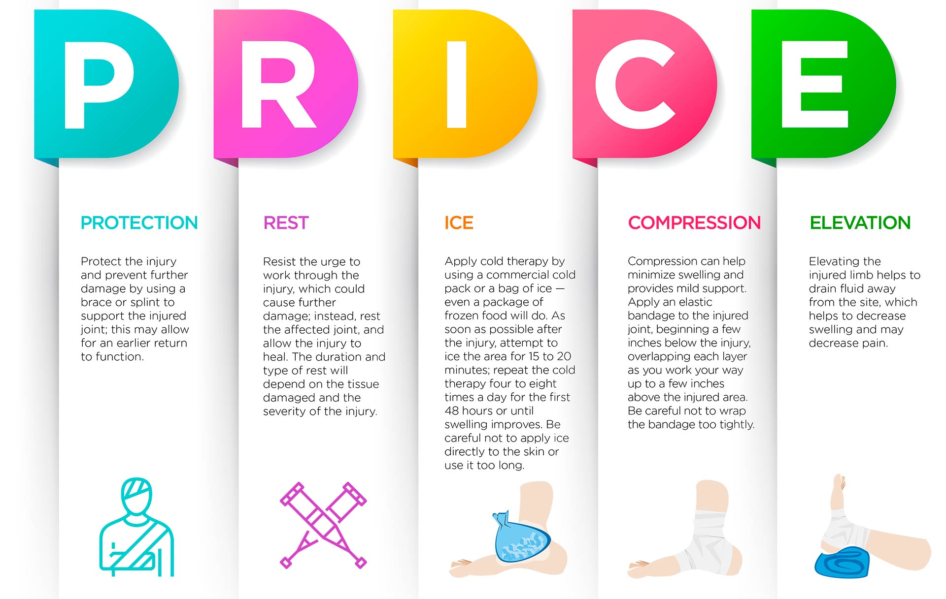
Sprains can be successfully managed by following the mnemonic PRICE: protection, rest, ice, compression and elevation.
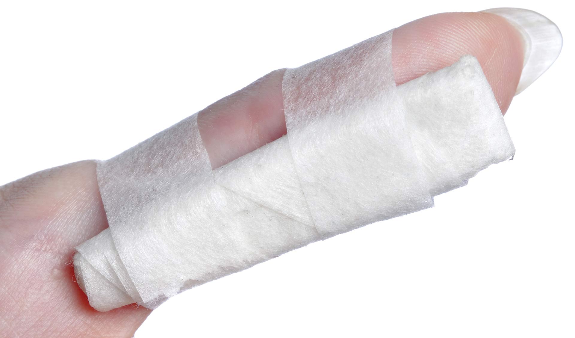
Protect the injury and prevent further damage by using a brace or splint to support the injured joint; this may allow for an earlier return to function.
Resist the urge to work through the injury, which could cause further damage; instead, rest the affected joint, and allow the injury to heal. The duration and type of rest will depend on the tissue damaged and the severity of the injury.
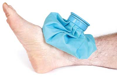
Apply cold therapy by using a commercial cold pack or a bag of ice — even a package of frozen food will do. As soon as possible after the injury, attempt to ice the area for 15 to 20 minutes; repeat the cold therapy four to eight times a day for the first 48 hours or until swelling improves. Avoid frostbite by being careful not to apply ice directly to the skin or use it too long.
Compression can help minimize swelling and provides mild support. Apply an elastic bandage to the injured joint, beginning a few inches below the injury, overlapping each layer as you work your way up to a few inches above the injured area. Be careful not to wrap the bandage too tightly.
Elevating the injured limb helps to drain fluid away from the site, which helps to decrease swelling and may decrease pain.
Use mild oral over-the-counter pain relievers and analgesics or topical ointments to lessen the pain. Nonsteroidal anti-inflammatory drugs (NSAIDs) can be taken orally (ibuprofen, naproxen, meloxicam) or applied directly to the skin (Pennsaid® or Voltaren® gel) overlying the painful joint.
Mild sprains may briefly impair one’s ability to perform regular activities on a boat, interfere with some sports practices and complicate travel. Severe injuries with deformity or complete loss of function require evacuation to the nearest medical facility.
To help prevent sprains, one should wear proper boating footwear — that is, with a good grip and a thin sole. Warm up properly before activities, gently stretch after warm-up, and do regular strengthening and flexibility exercises.

A dislocation occurs when the bones that articulate at a joint are forced out of position and no longer work closely each other in normal alignment. The separation of the bones, which may be complete (dislocation) or partial (subluxation), results from trauma or an extreme position that overstresses the joint.
The most common dislocations in adults occur in the shoulder and fingers. Any joint can be affected, including the hip, knee, ankle or jaw. In children, the most common dislocation is of the elbow.
Pain may be moderate or severe and is accompanied by swelling and deformity. The victim may experience numbness and/or tingling at the joint or beyond it. There is often discoloration, bruising and an inability to move the joint. It can be difficult to distinguish between a dislocation and a fracture, and it will usually require an X-ray or other medical imaging to accurately diagnose the condition.
Dislocations are orthopedic emergencies that require treatment in a medical facility and often require sedation or deep anesthesia to relocate the joint. Until medical help is available, splint the affected joint as it lies in its fixed position. Do not try to move a dislocated joint or force it back into place. This can damage the joint and its surrounding muscles, ligaments, nerves or blood vessels.
Dislocations require treatment by a medical professional. Do not delay medical care.
Avoid standing on unstable objects. Employ safe lifting mechanics, and wear supportive, nonslip shoes. Avoid hyperextension of the fingers.

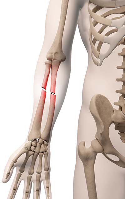
A fracture is a break in the bone. Various mechanisms cause fractures, so one needs to be aware that other injuries may be associated with the fracture. Obvious causes such as falls or significant impacts are readily associated with injury, but subtle twists or shifts that can occur aboard a boat can cause a fracture in older adults and in others with underlying medical conditions.
Fractures can be open (compound) or closed (simple). Open fractures are those in which the bone is exposed through the skin through either an open wound or a break in the skin. Closed fractures are those in which the bone does not penetrate the skin. Permanent injury to an extremity and serious (sometimes chronic) bone infection can result from an open fracture.
Medical imaging is used to diagnose a fracture. That technology will not be available until the injured person is evacuated to a medical facility that has those capabilities, so you will need to rely on a physical exam. Be aware that there may be other serious, subtler injuries, especially if significant trauma is involved. As with any trauma, remember the mnemonic ABCDE: airway, breathing, circulation, disability and exposure/environment. Rule out shock, and remember fractures can be associated with significant bleeding even if the fracture site is closed such as with the thigh or pelvis.
Unusual angulation of the bone, bone penetrating through the skin or a new “joint” that allows movement in a previously straight rigid bone can be apparent. More subtle symptoms may be tenderness at a specific point along the bony structure, slight angulation/shortening of the extremity, grinding sensation with any movement limb, inability to bear any weight, rapid bruising or bleeding at the site of injury and wound overlying the site of injury.
To determine if there is any damage to nerves and/or blood vessels, test to see if there is loss of sensation or motor function by employing many of the same techniques used in a neurological exam. A lack of pulses distally or an extremity that has become pale and cold in comparison with the other limb may signify vascular impairment. If you find any significant neurovascular deficits, intervention is imperative because permanent limb injury may occur in as little as four to six hours, depending on the degree of neurovascular injury.
Remove the victim from any environment that could endanger the victim or the rescuers. Employ trauma ABCDE protocols:
To remove clothing, cut away garments to prevent unnecessary movement of the fractured limb.
If you suspect neurovascular compromise, promptly but carefully realign the extremity enough to restore blood flow, which can be confirmed by palpable pulses. Traction may be necessary to obtain the desired result; it may also decrease the level of pain once the extremity is better aligned.
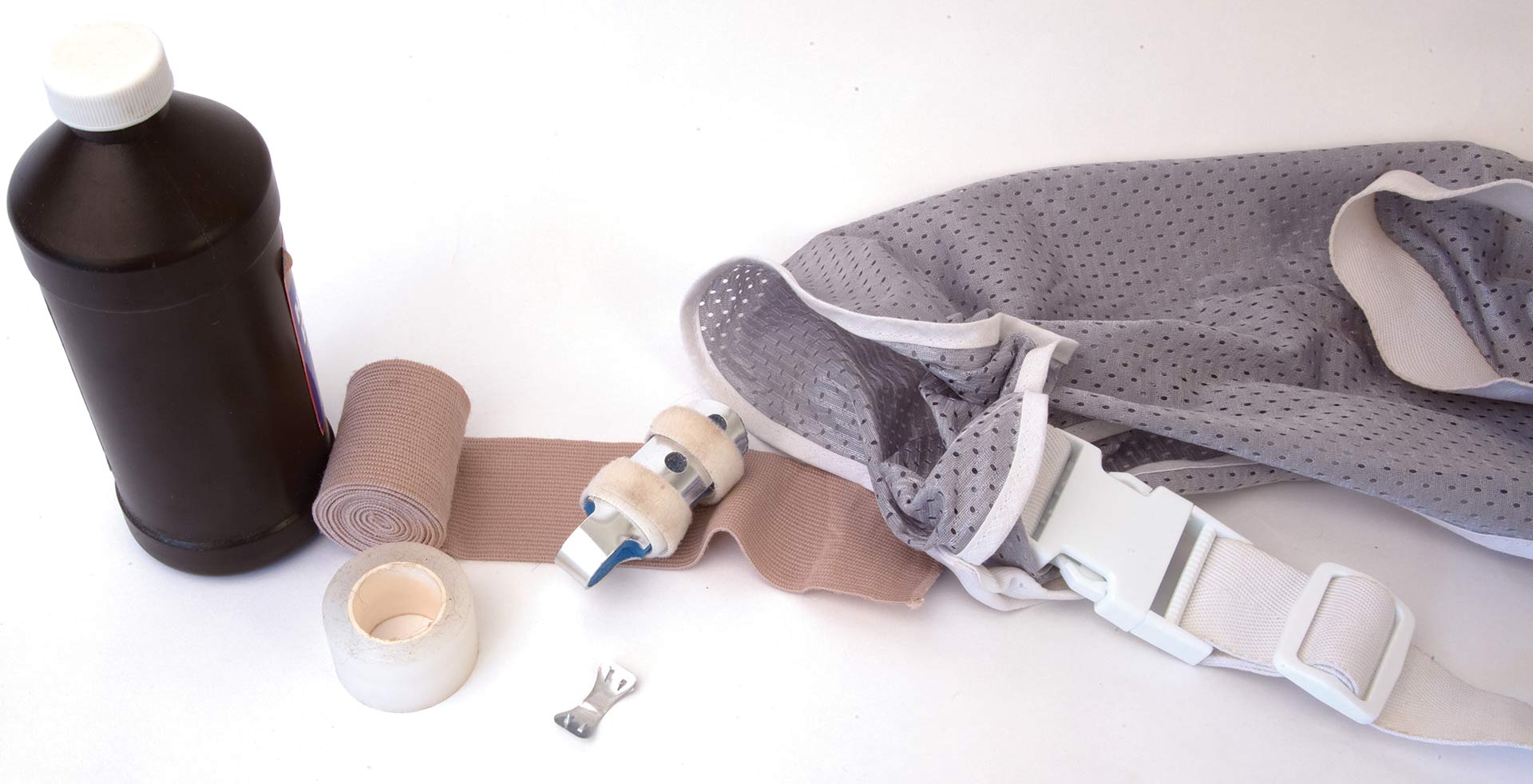
Splinting the fracture will be necessary to maintain the alignment, prevent additional injury from contact of the broken bony surfaces with surrounding tissue, decrease pain and facilitate transport. You can make a splint from available material that provides rigid support through the joint above and below the fracture and secures this support without compromising blood flow. Excellent splints are available commercially that are compact and easily carried in the event of such injuries.
Open fractures pose a serious infection risk. Often only a small wound overlies the fracture site. Assume that is the location where the bone fragment penetrated the skin and became contaminated with bacteria-laden debris before returning below the skin. Attempt to clear debris from the wound, preferably with copious sterile flushing, and apply a sterile dressing, if available. Cover the wound with an appropriate antibiotic, if available.
Evacuation is important, especially with open fractures or neurovascular compromise. During transport, regularly assess the victim’s neurovascular status. Once any wounds are dressed and bleeding is stopped, there is no need to reexplore until the victim reaches a medical facility for more definitive care.
Determine tetanus immunization status, record time of any tourniquet application, administer antibiotics and pain medicine if available, and record vital signs/neurovascular status at regular intervals in transit.
Be cognizant of fall or trip hazards. As important, identify your current position with the location of adequate medical facilities before conducting a hazardous operation that could be conducted later or with better equipment or more assistance. Employ additional safety measures if the need to proceed is determined.
An ill-defined mental condition called “vacationmania” appears to affect many people when they are on vacation in a spectacular location — where they disregard their normal reservations and concerns and instead feel invincible. They may try a range of risk-taking adventures, ranging from food to transportation to recreation. This happens to most people to some extent; just be aware of it as it is especially pertinent to traumatic injury prevention.
Reconsider renting motor vehicles — especially mopeds, motorcycles, etc. — if you are unfamiliar with their operation or with the area in which you are attempting to operate them. Maintenance of the equipment, different driving rules or customs, and questionable EMS capabilities are worthy of your serious consideration as well.
Include adequate supplies such as splints, irrigation, dressing supplies and antibiotics in your first-aid kit. Ensure that your tetanus immunization is current before traveling.
A fracture, especially open or with neurovascular compromise, is an emergency that will require the evacuation of the victim and usually the loss of his or her services to the expedition. The victim should avoid diving or other aquatic activities until the fracture has healed and a physician has cleared the victim for activity.
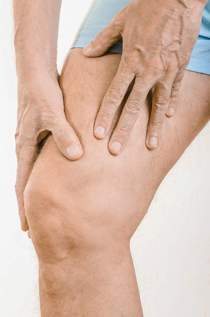
Tendonitis (or tendinitis) is inflammation of a tendon, which is a strong, flexible cordlike structure that connects a muscle to the bone. It is commonly caused by prolonged repetitive activity leading to overuse of a particular body part. It can also occur as a result of trauma or external compression.
Common locations of tendonitis include the elbow (tennis elbow), ankle (Achilles tendinitis), wrist (de Quervain’s tenosynovitis), knee (patellar tendonitis or jumper’s knee) and shoulder (rotator cuff tendonitis or bicipital tendonitis).
Pain with use of the affected muscle-tendon unit is a hallmark of tendonitis. The acute pain, which is usually located close to a joint, makes it difficult to move the affected joint. Weakness usually accompanies the pain.
Tenderness directly over the affected tendon will be present, often with mild swelling. If the affected tendon passes through a sheath (a fibrous tube that surrounds some tendons), crepitus (painful grinding, crackling or popping) may be experienced.
Tendonitis can usually be successfully managed by following the mnemonic RICE: relative rest, ice, compression and elevation.
An extended period of complete rest is no longer advocated, because it can lead to disuse atrophy (muscle shrinkage or weakness) and joint stiffness. A short period of immobilization for the affected area with early return to function is preferred, but avoid the provocative activity that caused the tendonitis. A brace or splint is often helpful to support the injured tendon and allows earlier return to function.
Use of mild pain relievers or analgesics such as acetaminophen (Tylenol®) taken orally or topically applied menthol (Biofreeeze® or similar) can lessen the pain.
NSAIDs can be taken orally (ibuprofen, naproxen, meloxicam) or applied directly to the skin (Pennsaid® or Voltaren® gel) overlying the painful area.
Severe cases of tendonitis may require formal physical therapy. Corticosteroids can be delivered to the site of inflammation via sterile needle injection or modalities using ultrasound (phonophoresis) or a small electric current (iontophoresis) to deliver the medication transcutaneously (through the overlying skin).
To help prevent tendonitis, avoid repetitive activities that can lead to overuse and the development of inflammation. Wear the correct footwear, warm up properly before activities, gently stretch after warm-up, and do regular strengthening and flexibility exercises.
Tendonitis may briefly impair one’s ability to perform regular activities on a boat and may complicate travel. Severe tendonitis may be disabling and require injection therapy.
Back pain will affect 80 percent of people over the course of their lives. Acute back pain may result from an injury to muscles, tendons, ligaments, facet joints, intervertebral discs, nerves or vertebrae of the thoracic (mid-back) or lumbar (lower back) area. Back pain can also result from injury to the sacroiliac (SI) joint, which connects the sacrum (tail bone) and the ilium (pelvis bone).
Most injuries that cause back pain occur as a result of improper or forceful lifting, but they can also occur as a result of a twisting injury, a misstep or a fall.
Back pain may also result from referred pain from nonorthopedic conditions such as kidney stones and infections, pancreatitis, ulcerative colitis, and other acute and potentially serious conditions.
Symptoms may be mild and aggravated by certain positions of sitting or standing, or they may be severe, incapacitating pain that prevents even basic functions such as standing erect or walking.
Back pain can be divided into two general types:
Mild back pain can often be alleviated by a short period of rest (less than 24 hours), avoiding strenuous lifting or pulling, minimizing bending and twisting, using ice and/or heat judiciously, gentle stretching and proper use of NSAIDs (ibuprofen, naproxen, meloxicam) or acetaminophen/paracetamol (Tylenol®).
Severe back or radicular pain will often require medication in the form of corticosteroids (prednisone), muscle relaxers and prescription pain medication.

Physical therapy, chiropractic care or manipulative therapy can also be beneficial. If symptoms do not improve within a few days, then it’s paramount to see an experienced medical provider for an examination to determine what is causing the back pain. The exam may include X-rays and an MRI.
Follow these suggestions to help prevent back pain:
Mild back pain may briefly impair one’s ability to perform regular activities on a boat and may complicate travel. Severe back injuries or worsening neuromuscular function require evacuation to the nearest hospital.
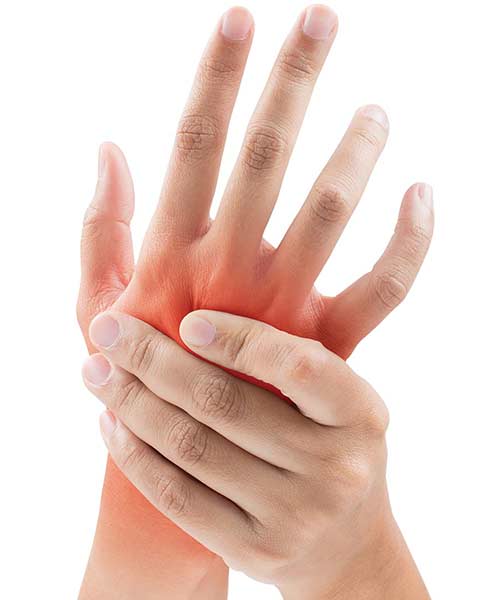
Joint pain (arthralgia) is a common condition that most people will experience at some point in their lifetime. Among the many causes of arthralgia are injury, infection, arthritis and systemic (whole body) illness, including allergic reaction. It can be experienced in any joint of the body, including the jaw, neck, shoulders, elbows, hands, hips, knees, ankles or feet.
Inflammatory arthralgia is distinguished by pain associated with joint swelling, heat and redness. It may be accompanied by systemic symptoms of fever, fatigue or weight loss.
Noninflammatory arthralgia is generally joint pain without significant swelling, heat, redness or systemic symptoms. An exception is a painful swollen joint that occurs after a trauma; if associated with joint deformity, it may suggest a fracture or dislocation.
Mild joint pain can usually be successfully managed by following the mnemonic RICE: relative rest, ice, compression and elevation. This method uses a multimodal approach to decrease the joint inflammation. Use of mild pain relievers or analgesics such as acetaminophen (Tylenol®) taken orally or topically applied menthol (Biofreeze® or similar) can lessen the pain.
NSAIDs can be taken orally (ibuprofen, naproxen, meloxicam) or applied directly to the skin (Pennsaid® or Voltaren® gel) overlying the painful joint. NSAIDs such as diclofenac or ketorolac can be purchased without prescriptions in some countries. While these drugs can be very effective, people with known gastrointestinal, cardiovascular, hepatic and renal issues should exercise caution when using them. Take these drugs only as instructed, and do not use them for more than a week at a time unless they were prescribed by a medical doctor.
It is not possible to prevent all causes of joint pain, but healthy lifestyle choices can reduce the risk. Follow these suggestions to help prevent or reduce the risk of arthralgia:
Mild joint pain may briefly impair one’s ability to perform regular activities on a boat and may complicate travel. Severe joint pain associated with trauma and deformity or progressive systemic illness requires evacuation to the nearest hospital.
The neck is remarkable: It supports the head and provides it with a wide range of mobility. It encompasses critical vascular structures such as the carotid arteries and jugular vein; neural structures such as cranial, peripheral nerves and spinal cord; gastrointestinal structures such as the esophagus; respiratory structures such as the trachea, etc. The complex musculature finds many of its attachments connected to the cervical spine. When the neck is subjected to trauma, all of these areas need to be evaluated properly.
Pain and muscle spasm are two symptoms that may indicate neck trauma has injured the neck. Significant injury can be found when motor weakness or paralysis is noted below the level of the injury. Reflex changes may occur when inhibitory signals are blocked. Arthritic joints in the neck may cause significant discomfort and decrease range of motion. Headaches can occur with chronic neck pain and spasm. A large study found the need for imaging was greatly reduced if the victim has normal alertness, no drugs or alcohol, no posterior neck tenderness, no other painful injuries and a normal neurological and mental status exam.
When neck injury is suspected, immobilization of the neck is recommended to avoid further injury to the neck and spinal cord in case the elements may have been injured and provide little or no protection. Place a cervical collar, if available, on the victim. Otherwise, block the head with towel rolls or foam blocks or even tape the head. Use of spine board may be helpful, but some medical professionals question its use, especially if a prolonged time is anticipated before receiving definitive care. Unstable neck injuries may require surgery. Fortunately, most injuries are self-limited and respond to conservative therapies.
If a significant injury is associated with neurological or motor deficits, evacuate the victim to a medical facility, preferably a clinic with imaging capability. Refrain from diving or other activities until any motor or sensory deficits have cleared. Lying in a horizontal position with the head up may worsen neck issues.
Take care to prevent slips and falls. Wear proper footwear, provide nonskid surfaces, ascend or descend steps deliberately and carefully while holding a railing when able, and pay attention to overhanging equipment such as booms, hatches, etc.
Muscle strain is a common term that encompasses injury to skeletal muscle. It is sometimes referred to as a pulled muscle. The injury occurs when a muscle is stretched to its limits or is explosively stretched too quickly. The muscle itself may actually tear on such strain.
The most common symptom is acute pain in the muscle groups that have been subjected to the strain. Weakness in the affected muscle group can result from the injury. Muscle is a heavily vascular tissue, so disruption of the blood vessels may result in bleeding into the muscle, creating a bruise in that area. Swelling is noted as the injured muscle becomes edematous (full of excess fluid) and swollen. The muscle may spasm and cause additional pain. A complete tear or avulsion (separation) of the muscle will result in pain, deformity and loss of motor function for that muscle group.
Apply ice to the area for several hours on the first day. This may lessen the swelling and bleeding that can occur with severe strains. Elevating the limb can decrease the swelling and pain by increasing the drainage of fluid and blood from that area. Some medical professionals find that wrapping the injured muscle can help with decreasing the pain and increasing function. Victims can use analgesics (pain relievers) judiciously if their pain is moderate or severe despite conservative measures. Progressive muscle rehabilitation — including stretching and exercise — helps to restore the muscle to its healthy baseline function.
To prevent muscle strain, gradually work up to the level of physical activity anticipated to be needed, with additional accounting for unexpected demand for even higher output. This process entails stretching, exercise and even weightlifting to increase needed strength for certain activities.
Diving with muscle strain can have a serious impact on the victim, especially if cramping or decreased motor activity secondary to pain occurs. Not being able to swim effectively from a hazardous situation or return to the surface can be life-threatening. Likewise, a significant strain that sidelines a boat’s crew member can have a significant impact on a vessel’s ability to function, provide personnel for its watches or perform its many daily functions. The possible need to divert others to assist the injured crew member can affect daily functions as well.
Since its inception in 1980, DAN® has spent more than 300,000 medical resource hours on duty, fielding more than 100,000 emergency calls and coordinating more than 10,000 emergency evacuations worldwide, making it one of the world’s most trusted resources for traveler safety.
As a natural evolution of DAN’s successful 35+ years of service to the diving community, our new DAN Boater Travel Safety Association is committed to helping recreational boaters and global travelers in need of emergency assistance while on the go, and to promoting smart travel health and safety practices through research and education.
By boat, plane, car, or rail. Around the world, or across the state. Anytime you travel more than 50 miles from home, rely on DAN Boater for the assistance you need when you need it:
Find out how our affordable annual travel safety memberships help travelers like you and your family every day.